41 ribosome diagram with labels
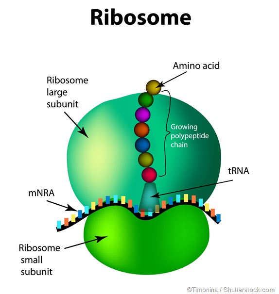
Solved In the following diagram of a ribosome, assign the | Chegg.com in the following diagram of a ribosome, assign the correct labels. 5' end of the mrna growing polypeptide a trna attached to a single amino acid ontors here large subunit atrna attached to a polypeptide is found in this area of the nibosome a trna that is not attached to anything exits hore 3' end of the mrna a trna moleculo mossenger rna being … Ribosome and protein synthesis, diagram - Science Photo Library Diagram showing protein synthesis in cells (translation). Messenger ribonucleic acid (mRNA, blue with coloured nucleotides) is read by a ribosome (pink). The molecules of transfer RNA (tRNA, key-shaped) each bring an amino acid (orange dot) to bind to the ribosome's protein synthesis site.
Mastering Biology Chapter 14 Flashcards - Quizlet Place the blue labels in their proper locations on this diagram showing the process of transcription. Then, use the pink labels to identify the corresponding RNA nucleotide that belongs in each pink target. Pink labels can be used once, more than once, or not at all. Drag the appropriate labels to their respective targets.
Ribosome diagram with labels
Bio 1113 - Unit 11 - Gene Expression Flashcards | Quizlet In the following diagram of a ribosome, assign the correct labels: Label 1: a tRNA attached to a polypeptide is found in this area of the ribosome Label 2: a tRNA attached to a single amino acid enters here Label 3: a tRNA that is not attached to anything exits here Label 4: a tRNA molecule Label 5: growing polypeptide Label 6: mRNA being ... Animal Cells: Labelled Diagram, Definitions, and Structure Animal Cells Organelles and Functions. A double layer that supports and protects the cell. Allows materials in and out. The control center of the cell. Nucleus contains majority of cell's the DNA. Popularly known as the "Powerhouse". Breaks down food to produce energy in the form of ATP. Animal Cell Diagram with Label and Explanation: Cell ... - Collegedunia Animal cell is a typical Eukaryotic cell enclosed by a plasma membrane containing nucleus and organelles which lack cell walls, unlike all other Eukaryotic cells. The typical cell ranges in size between 1-100 micrometers. The lack of cell walls enabled the animal cells to develop a greater diversity of cell types.
Ribosome diagram with labels. DNA Labeling: Transciption and Translation - The Biology Corner This worksheet shows a diagram of transcription and translation and asks students to label it; also includes questions about the processes. Name: _____ ... How does the ribosome know the sequence of amino acids to build? 12. What is the difference between a codon and an anticodon? Prokaryotic Cells - BioNinja Prokaryotes are organisms whose cells lack a nucleus ('pro' = before ; 'karyon' = nucleus). They belong to the kingdom Monera and have been further classified into two distinct domains: Archaebacteria – found in extreme environments like high temperatures, salt concentrations or pH (i.e. extremophiles); Eubacteria – traditional bacteria including most known pathogenic forms … Cell Organelles- Definition, Structure, Functions, Diagram In the case of prokaryotic cells, the ribosomes are of the 70S with the larger subunit of 50S and the smaller one of 30S. Eukaryotic cells have 80S ribosomes with 60S larger subunit and 40S smaller subunit. Ribosomes are short-lived as after the protein synthesis, the subunits split up and can be either reused or remain broken up. Oxford Cambridge and RSA Friday 16 October 2020 – Morning ribosome Fig. 1.2. 3 ... State three changes, other than to the labels, to Fig. 1.2 that the student would need to ... Below is a diagram of a goblet cell as seen under an electron microscope. A B (i) Suggest why goblet cells have large numbers of the cellular component labelled A. ...
Plant Cell Diagram | Science Trends A plant cell diagram, like the one above, shows each part of the plant cell including the chloroplast, cell wall, plasma membrane, nucleus, mitochondria, ribosomes, etc. A plant cell diagram is a great way to learn the different components of the cell for your upcoming exam. Plants are able to do something animals can't: photosynthesize. ProteInfer - GitHub Pages Below we predict the GO terms of the protein. Gene Ontology (GO) describe the molecular function and biological processes that proteins take part in. GO terms are often experimentally validated, but there are a lot of unannotated proteins. GO terms have a hierarchical structure, from most general to most specific. In summary mode, on the left, we present the most specific … Mastering Biology Chapter 14 Flashcards - Quizlet Place the blue labels in their proper locations on this diagram showing the process of transcription. Then, use the pink labels to identify the corresponding RNA nucleotide that belongs in each pink target. Pink labels can be used once, more than once, or not at all. Drag the appropriate labels to their respective targets. Label Transcription and Translation - 7355418.pdf - Course Hero View Label Transcription and Translation - 7355418.pdf from BIOLOGY 101 at Harmony School of Innovation Fort Worth. 1. Label the diagram. DNA Protein Amino Acid Ribosome mRNA Codon tRNA Anticodon 2.
Ribosome - Wikipedia Prokaryotic ribosomes are around 20 nm (200 Å) in diameter and are composed of 65% rRNA and 35% ribosomal proteins. Eukaryotic ribosomes are between 25 and 30 nm (250-300 Å) in diameter with an rRNA-to-protein ratio that is close to 1. Nuclear envelope - Wikipedia The nuclear envelope is punctured by around a thousand nuclear pore complexes, about 100 nm across, with an inner channel about 40 nm wide. The complexes contain a number of nucleoporins, proteins that link the inner and outer nuclear membranes.. Cell division. During the G2 phase of interphase, the nuclear membrane increases its surface area and doubles its … Ribosomes Stock Illustrations - 641 Ribosomes Stock ... - Dreamstime Download 641 Ribosomes Stock Illustrations, Vectors & Clipart for FREE or amazingly low rates! New users enjoy 60% OFF. 187,336,869 stock photos online. ... Anatomical and medical labeled scheme. Explained closeup diagram. Ribosomes vector illustration. Anatomical and medical labeled. Ribosomes- Definition, Structure, Functions and Diagram Ribosomes Definition The ribosome word is derived - 'ribo' from ribonucleic acid and 'somes' from the Greek word 'soma' which means 'body'. Ribosomes are tiny spheroidal dense particles (of 150 to 200 A0 diameters) that are primarily found in most prokaryotic and eukaryotic. They are sites of protein synthesis.
Campbell Ap Biology Mastering Biology Chapter 17 Course Work Drag the white and purple labels to the white targets to indicate what each mutant mRNA codon codes for. (You will probably need to consult the codon table for mRNA .) Drag the pink labels to the pink targets to indicate the type of mutation. Drag the blue labels to the blue targets to indicate the effect on the polypeptide's primary structure.
Cambridge Assessment International Education Cambridge 1 The diagram shows a leaf on a plant. Sun water from the soil carbon dioxide from the air simple sugars made in the leaf Which characteristic of life is represented by this diagram? A excretion B nutrition C respiration D sensitivity 2 The diagram shows how Homo sapiens (modern people) could have evolved from earlier ancestors. Homo habilis ...
PDF Quick Review Transcription and Translation - WPMU DEV label the diagram. 2. ... 910dnamrnait carries the genetic code from dna to ribosome to make a proteinit carries the amino acids to make proteinbecause the genetic code is the recipe to make a protein and is contained in a mrnacodons are in mrna and anti codons are groups of 3 bases in trnatranscription takes place in nucleus; translation takes ...
Solved The ribosome in the diagram is in the process of | Chegg.com The ribosome in the diagram is in the process of synthesizing a protein using directions transcribed from the DNA. Use the labels to identify each of the structures involved in translation and protein synthesis. Question: The ribosome in the diagram is in the process of synthesizing a protein using directions transcribed from the DNA.
Biology: parts of a ribosome Diagram | Quizlet Start studying Biology: parts of a ribosome. Learn vocabulary, terms, and more with flashcards, games, and other study tools.
ProteInfer - GitHub Pages We focus on Swiss-Prot to ensure that our models learn from human-curated labels, rather than labels generated by a computational annotation pipeline. Each protein in Swiss-Prot goes through a 6-stage process of sequence curation, sequence analysis, literature curation, family-based curation, evidence attribution, and quality assurance.
Structure of Ribosome - Biology Wise Diameter of Ribosome is 20nm. Their composition can be divided into two parts - 2/3 part of r-RNA (ribosomal RNA) and 1/3 part RNP (Ribosomal protein or Ribonuclep protein). Polypeptide chain is fabricated by translating mRNA (messenger RNA) with the aid amino acids that tRNA (transfer RNA) delivers.

FREE, gadget, Diagram of a ribosome #BacktoSchoolWithVersal | Cell structure, Cell, Science projects
Nuclear envelope - Wikipedia The nuclear envelope is punctured by around a thousand nuclear pore complexes, about 100 nm across, with an inner channel about 40 nm wide. The complexes contain a number of nucleoporins, proteins that link the inner and outer nuclear membranes.
GRADE 12 LIFE SCIENCES LEARNER NOTES - Mail & Guardian 1.1 The diagram below represents a part of a molecule. Study the diagram and answer the questions that follow. 1.1.1 Identify the molecule in the above diagram. (1) 1.1.2 Label the parts numbered 1 and 5 respectively. (2) 1.1.3 What is the collective name …
What Are Ribosomes? - Definition, Structure and its Functions Ribosomes are located inside the cytosol found in the plant cell and animal cell. The ribosome structure includes the following: It is located in two areas of cytoplasm. Scattered in the cytoplasm. Prokaryotes have 70S ribosomes while eukaryotes have 80S ribosomes. Around 62% of ribosomes are comprised of RNA, while the rest is proteins.
Protein Targeting (With Diagram) | Molecular Biology ADVERTISEMENTS: Let us make an in-depth study of the protein targeting. After reading this article you will learn about: 1. Introduction to Protein Targeting 2. Signal Sequence 3. Transport of Proteins into ER 4. Signal Sequence Recognition Mechanism 5. Role of Golgi Complex in Protein Transportation 6. Transport of Proteins from Golgi to Lysosomes 7. […]
Active Ribosome Profiling with RiboLace - PubMed Ribosome profiling, or Ribo-seq, is based on large-scale sequencing of RNA fragments protected from nuclease digestion by ribosomes. Thanks to its unique ability to provide positional information about ribosomes flowing along transcripts, this method can be used to shed light on mechanistic aspects … Active Ribosome Profiling with RiboLace
Structure of Ribosome (With Diagram) - Biology Discussion A bacterial ribosome is about 250 nm in diameter and consists of two subunits, one large and one small. Both subunits consist of one or more molecules of rRNA and an array of ribosomal proteins. ADVERTISEMENTS: Association of two subunits is called mono-some. The structure of prokaryotic ribosome is given in the figure 8.2 B.
GRADE 12 LIFE SCIENCES LEARNER NOTES - Mail & Guardian 1.1 The diagram below represents a part of a molecule. Study the diagram and answer the questions that follow. 1.1.1 Identify the molecule in the above diagram. (1) 1.1.2 Label the parts numbered 1 and 5 respectively. (2) 1.1.3 What is the collective name for the parts numbered 2, 3 and 4? (1)
Ribosome - protein factory - definition, function, structure and biology The protein translation by a ribosome consists of three stages: (1) Initiation, (2) Elongation, and (3) Termination. Initiation - the ribosome assembles around the target mRNA. A small ribosome subunit links onto the "start-end" of an mRNA strand. "Initiator tRNA" also enters the small subunit and binds to the start codon (most commonly, AUG).


Post a Comment for "41 ribosome diagram with labels"