38 cell diagram and labels
en.wikipedia.org › Cell_membrane_(diagrammatic)Wikipedia:Featured picture candidates/Cell membrane ... Edit 3 uploaded Standard zoom boxes tend to obscure the labels, but if there is a consensus to change to those, I'll give it another try. Dhatfield ( talk ) 20:29, 16 June 2008 (UTC) [ reply ] Strong oppose There are only unsaturated tails on the phospholipids, for a reduced structure diagram of a cell membrane's lipid bilayer there should be ... label diagram of tissue cells Plant tissues. label diagram of tissue cells. Basic Histology -- Smooth Muscle, Longitudinal Section we have 9 Pics about Basic Histology -- Smooth Muscle, Longitudinal Section like Pseudostratified Ciliated Columnar Epithelium Shows Cilia Ciliated, Human Anatomy Lab Exercises Tissues Recognition and Function Flashcards and also Nervous Tissue ...
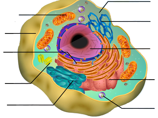
Animal Cells: Labelled Diagram, Definitions, and Structure The endoplasmic reticulum (s) are organelles that create a network of membranes that transport substances around the cell. They have phospholipid bilayers. There are two types of ER: the rough ER, and the smooth ER. The rough endoplasmic reticulum is rough because it has ribosomes (which is explained below) attached to it.

Cell diagram and labels
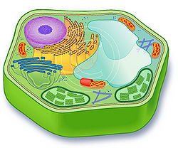
Plant Cells Vs. Animal Cells (With Diagrams) - Owlcation Large Vacuole: While animal cells may have many tiny vacuoles, a plant cell usually has a single large vacuole, which serves as a storage tank for food, water, waste products, and other materials. The vacuole has an important structural function, as well. When filled with water, the vacuole exerts internal pressure against the cell wall, which ... 03 Label the Cell Diagram | Quizlet Cell Biology 03 Label the Cell STUDY Learn Flashcards Write Spell Test PLAY Match Gravity Created by muskopf1TEACHER Terms in this set (14) Nucleus Control center of the cell Nucleolus Ribosome synthesis Rough Endoplasmic Reticulum Protein transport Smooth Endoplasmic Reticulum Lipid synthesis Mitochondrion Cellular Respiratoin Golgi Apparatus Cell: Structure and Functions (With Diagram) - Biology Discussion Eukaryotic Cells: 1. Eukaryotes are sophisticated cells with a well defined nucleus and cell organelles. 2. The cells are comparatively larger in size (10-100 μm). 3. Unicellular to multicellular in nature and evolved ~1 billion years ago. 4. The cell membrane is semipermeable and flexible. 5. These cells reproduce both asexually and sexually.
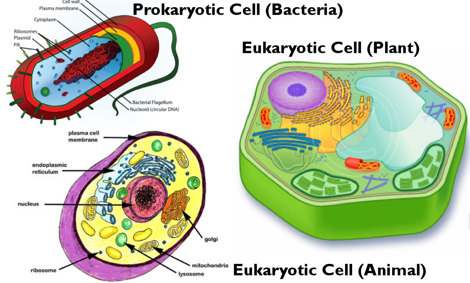
Cell diagram and labels. A Labeled Diagram of the Plant Cell and Functions of its Organelles A Labeled Diagram of the Plant Cell and Functions of its Organelles We are aware that all life stems from a single cell, and that the cell is the most basic unit of all living organisms. The cell being the smallest unit of life, is akin to a tiny room which houses several organs. Here, let's study the plant cell in detail... A Labeled Diagram of the Animal Cell and its Organelles A Labeled Diagram of the Animal Cell and its Organelles There are two types of cells - Prokaryotic and Eucaryotic. Eukaryotic cells are larger, more complex, and have evolved more recently than prokaryotes. Where, prokaryotes are just bacteria and archaea, eukaryotes are literally everything else. Free Cell Diagram Software with Free Templates - EdrawMax An animal cell diagram describes a cell structure enclosed by a plasma member, and it has a nucleus with a membrane and organelles. Neuron Diagram A neuron diagram describes the three parts of a Neuron: dendrites, an axon, a cell body, or soma. Cell Membrane Diagram Plant Cell Diagram | Science Trends A plant cell diagram, like the one above, shows each part of the plant cell including the chloroplast, cell wall, plasma membrane, nucleus, mitochondria, ribosomes, etc. A plant cell diagram is a great way to learn the different components of the cell for your upcoming exam. Plants are able to do something animals can't: photosynthesize.
Animal Cell Diagram PDF in 2022 - Pinterest Budding biologists, here's a great cheat-sheet to help you memorize and compare the structures of both animal cells and plant cells. A printable diagram of an animal cell. This PDF includes the color version, black and white version, and the labeled and unlabeled diagrams for students to complete. 6 pages total. Cell Organelles- Definition, Structure, Functions, Diagram An additional non-living layer present outside the cell membrane in some cells that provides structure, protection, and filtering mechanism to the cell is the cell wall. Structure of Cell Wall. In a plant cell, the cell wall is made up of cellulose, hemicellulose, and proteins while in a fungal cell, it is composed of chitin. A cell wall is ... Plant Cells: Labelled Diagram, Definitions, and Structure Plastids and Chloroplasts. Plants make their own food through photosynthesis. Plant cells have plastids, which animal cells don't. Plastids are organelles used to make and store needed compounds. Chloroplasts are the most important of plastids. They convert light energy from the sun into sugar and oxygen. The most exposed parts of the plants ... Bacteria in Microbiology - shapes, structure and diagram Bacterial spores. Bacterial endospores layers. Bacteria cells are the smallest living cells that are known; even though viruses are smaller than bacteria, viruses are not living cells. There are different types of bacteria with various sizes, shapes, and structures. The bacteria shapes, structure, and labeled diagrams are discussed below.
A Labelled Diagram Of Neuron with Detailed Explanations Diagram Of Neuron. A neuron is a specialized cell, primarily involved in transmitting information through electrical and chemical signals. They are found in the brain, spinal cord and the peripheral nerves. A neuron is also known as the nerve cell. The structure of a neuron varies with their shape and size and it mainly depends upon their ... Learn the parts of a cell with diagrams and cell quizzes Cell diagram unlabeled It's time to label the cell yourself! As you fill in the cell structure worksheet, remember the functions of each part of the cell that you learned in the video. Doing this will help you to remember where each part is located. Click the links below to download the labeled and unlabeled eukaryotic cell diagrams. Animal And Plant Cell Diagram To Label Worksheets & Teaching Resources ... Plant and Animal Cell Labeling Diagrams by A-Thom-ic Science 12 $2.00 PDF Three versions of the plant cell worksheet and three versions of the animal cell worksheet allow students of different grade levels and/or skill levels to label and review the parts of each cell type. Can be used as homework or a quiz. Cells Diagram | Science Illustration Solutions - Edrawsoft Cells Diagram Symbols Edraw software offers you lots of symbols used in cells diagram like cell structure, paramecium, squamous cell, cell division, bacteria, cell membrane, eggs, sperm, zygote, an animal cell, SARS, tobacco mosaic, adenovirus, coliphage, herpesvirus, AIDS, pollen, plant cell model, onion tissue, etc. Cells Diagram Examples
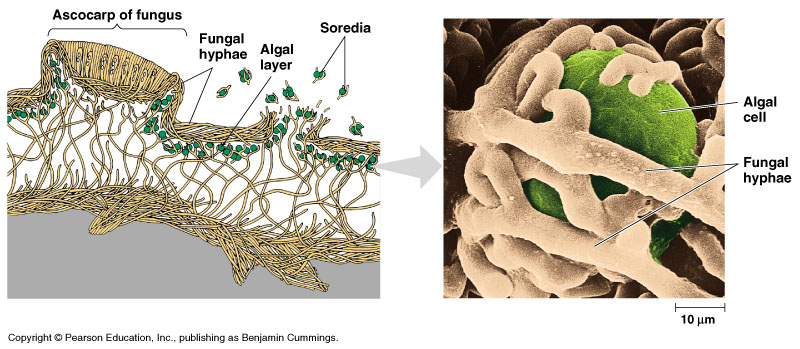
Fungi
Labeled Plant Cell With Diagrams | Science Trends The parts of a plant cell include the cell wall, the cell membrane, the cytoskeleton or cytoplasm, the nucleus, the Golgi body, the mitochondria, the peroxisome's, the vacuoles, ribosomes, and the endoplasmic reticulum. Parts Of A Plant Cell The Cell Wall Let's start from the outside and work our way inwards.
Quia - Meiosis Illustration Identification
Label Cell Parts | Plant & Animal Cell Activity | StoryboardThat Student Instructions Create a cell diagram with each part of plant and animal cells labeled. Include descriptions of what each organelle does. Click "Start Assignment". Find diagrams of a plant and an animal cell in the Science tab. Using arrows and Textables, label each part of the cell and describe its function.
Cell Diagram To Label - ClipArt Best
A Well-labelled Diagram Of Animal Cell With Explanation - Byju's The animal cell diagram is widely asked in Class 10 and 12 examinations and is beneficial to understand the structure and functions of an animal. A brief explanation of the different parts of an animal cell along with a well-labelled diagram is mentioned below for reference. Also Read Different between Plant Cell and Animal Cell

label diagram of cell
Animal Cell Labeled Diagram Pictures, Images and Stock Photos Browse 19 animal cell labeled diagram stock photos and images available, or start a new search to explore more stock photos and images. Newest results. Diagrams of animal and plant cells. Labelled diagrams of typical animal and plant cells with editable layers. Golgi apparatus or Golgi body.
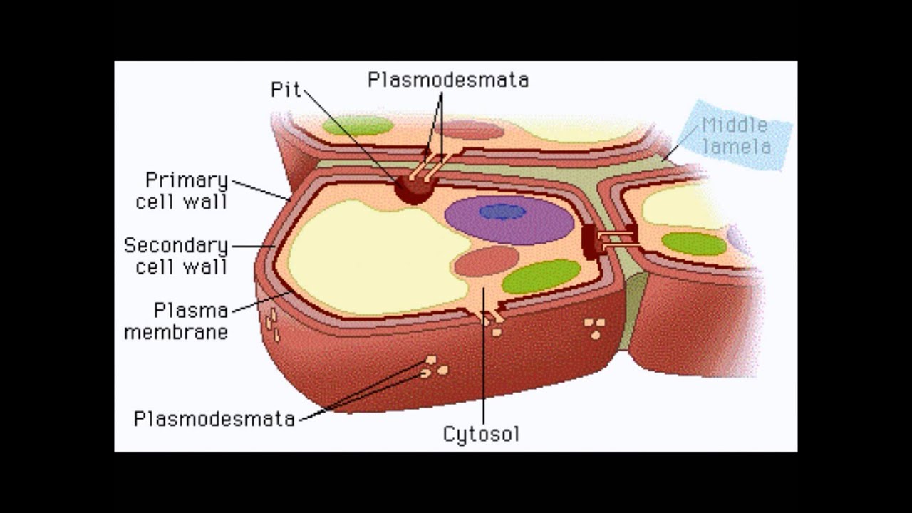
AP Biology - Plant Cell Wall - YouTube
IXL | Plant cell diagrams: label parts | 8th grade science Improve your science knowledge with free questions in "Plant cell diagrams: label parts" and thousands of other science skills.

A typical cell, labeled diagram. | Alila Medical Images
CELL MEMBRANE LABEL Diagram | Quizlet Practice labeling the parts of the cell membrane Terms in this set (6) Channel Protein hole or tunnel that particles may pass through to go in / out of cell Marker protein identifies or labels the cell Receptor protein receives information Heads part of the phospholipid that loves water (hydrophili) - points to the most outside and inside of cell
CELL - Labelled diagram
Plant and Animal Cell: Labeled Diagram, Structure, Function - Embibe Plant Cell: Plant cells are eukaryotic cells with a true nucleus along with specialized structures called organelles that carry out certain specific functions. Animal Cell: An animal cell is a type of eukaryotic cell that lacks a cell wall and has a true, membrane-bound nucleus along with other cellular organelles. Diagram of Plant and Animal Cell
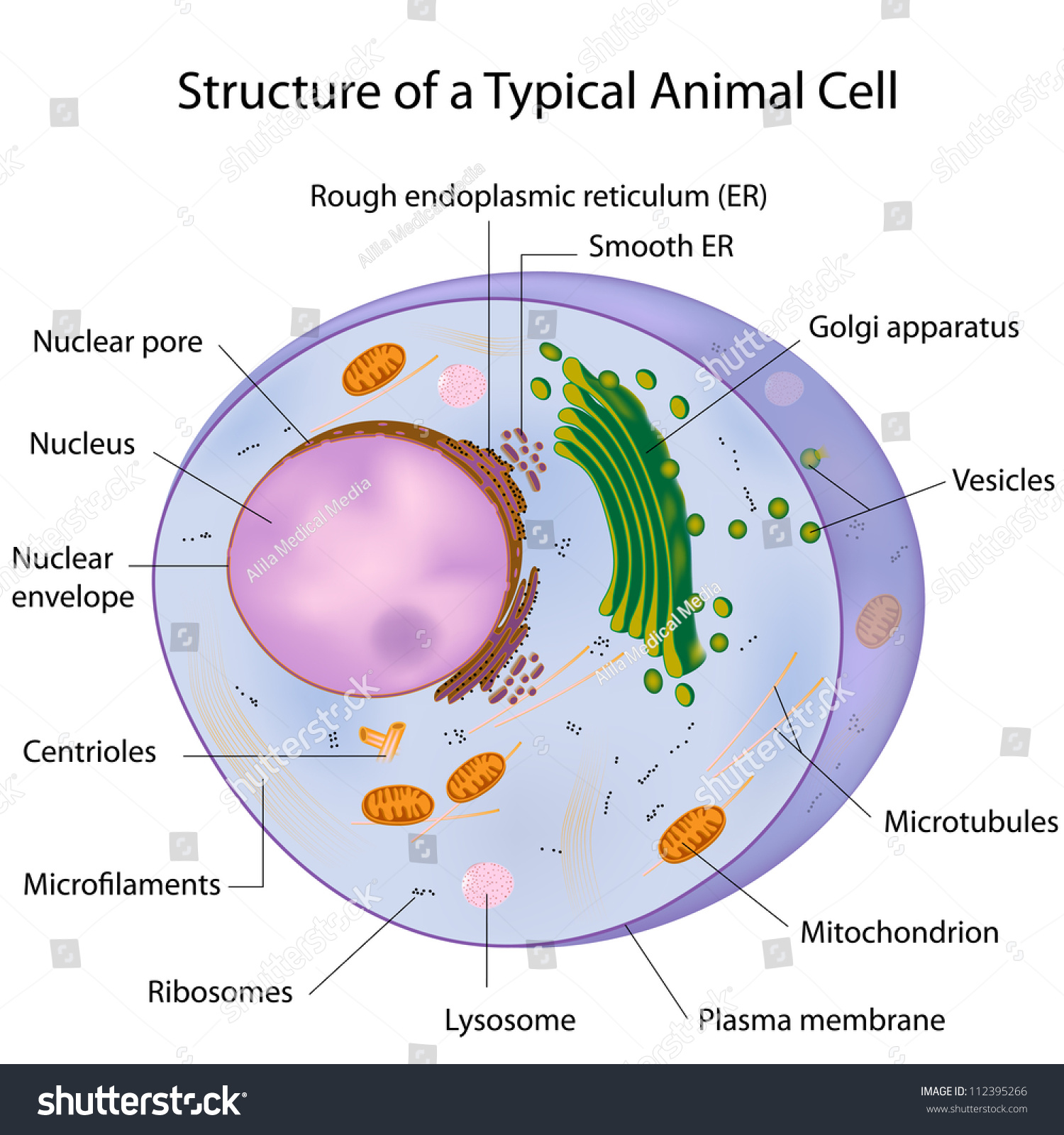
Typical Cell Labeled Stock Illustration 112395266 - Shutterstock
Animal Cell Diagram with Label and Explanation: Cell Structure, Functions Diagram of Animal Cell Below is the diagram of the animal cell which shows the organelles present in it. The cell is covered with cytoplasm which consists of cell organelles in it. The nucleus is covered with a rough Endoplasmic Reticulum and other organelles each designed for a specific purpose.

Label A Cell Diagram - Human Anatomy
Label that Diagram - Cells by rtcgames Label that Diagram - Cells This app provides the user the opportunity to study cell structure as well as test what they know. There are 5 cells presented: Animal Cell, Plant Cell, Amoeba, Paramecium and the Euglena. The player can study the labeled diagram or play the game of labeling the diagram. The app was created with Clickteam Multimedia ...

2944f5bade4b5af1aed64278d9d4db3f.jpg (236×236) | Animal cell project, Animal cell model project ...
Interactive Cell Model - CELLS alive Cell Wall. Chloroplast. Smooth Endoplasmic Reticulum. Rough Endoplasmic Reticulum. Ribosomes. Cytoskeleton. RETURN to CELL DIAGRAM ...
Print Exercise 7 flashcards | Easy Notecards
PDF Human Cell Diagram, Parts, Pictures, Structure and Functions One of the few cells in the human body that lacks almost all organelles are the red blood cells. The main organelles are as follows : cell membrane endoplasmic reticulum Golgi apparatus lysosomes mitochondria nucleus perioxisomes microfilaments and microtubules 2
Plant cell - SignWiki
Cell: Structure and Functions (With Diagram) - Biology Discussion Eukaryotic Cells: 1. Eukaryotes are sophisticated cells with a well defined nucleus and cell organelles. 2. The cells are comparatively larger in size (10-100 μm). 3. Unicellular to multicellular in nature and evolved ~1 billion years ago. 4. The cell membrane is semipermeable and flexible. 5. These cells reproduce both asexually and sexually.

My Classroom
03 Label the Cell Diagram | Quizlet Cell Biology 03 Label the Cell STUDY Learn Flashcards Write Spell Test PLAY Match Gravity Created by muskopf1TEACHER Terms in this set (14) Nucleus Control center of the cell Nucleolus Ribosome synthesis Rough Endoplasmic Reticulum Protein transport Smooth Endoplasmic Reticulum Lipid synthesis Mitochondrion Cellular Respiratoin Golgi Apparatus

Attack of The Silverfish!: Images of the stinging cells of Cnidaria
Plant Cells Vs. Animal Cells (With Diagrams) - Owlcation Large Vacuole: While animal cells may have many tiny vacuoles, a plant cell usually has a single large vacuole, which serves as a storage tank for food, water, waste products, and other materials. The vacuole has an important structural function, as well. When filled with water, the vacuole exerts internal pressure against the cell wall, which ...
Cell Diagram To Label - ClipArt Best
Biology - Ms. Ryalls... Midvale Science
![[DIAGRAM] Printable Animal Cell Diagram With Labels And Functions FULL Version HD Quality And ...](https://media.proprofs.com/images/QM/user_images/2503852/1573534103.jpg)
[DIAGRAM] Printable Animal Cell Diagram With Labels And Functions FULL Version HD Quality And ...
Parts and Function of Digestive System for Med School & Nursing Students - NCLEX Quiz
Post a Comment for "38 cell diagram and labels"