38 diagram of a human cell with labels
Label a human cell diagram - BonifaceWoods's blog The Cell - Courses Pages Label a human cell diagram The Cell - Courses Pages The Cell - Courses Pages Diagram Label Sperm Biology - vector clip art online, royalty free. a human muscle cell labeling worksheet - Oxycodon last longer in bed The Cell - Courses Pages File:Diagram human cell nucleus tr.svg - Wikimedia Commons Biology Quiz : Animal ... Best way to learn: Labeled Diagram Of A Human Blood Cell Labels that inform both medical professionals and consumers that these products were made possible by the taking of innocent human life. Diagram of the brain of a person The problem comes about due to the blood-brain barrier—natures way of preventing pathogens from getting into the central nervous system—its a layer of cells that line the ...
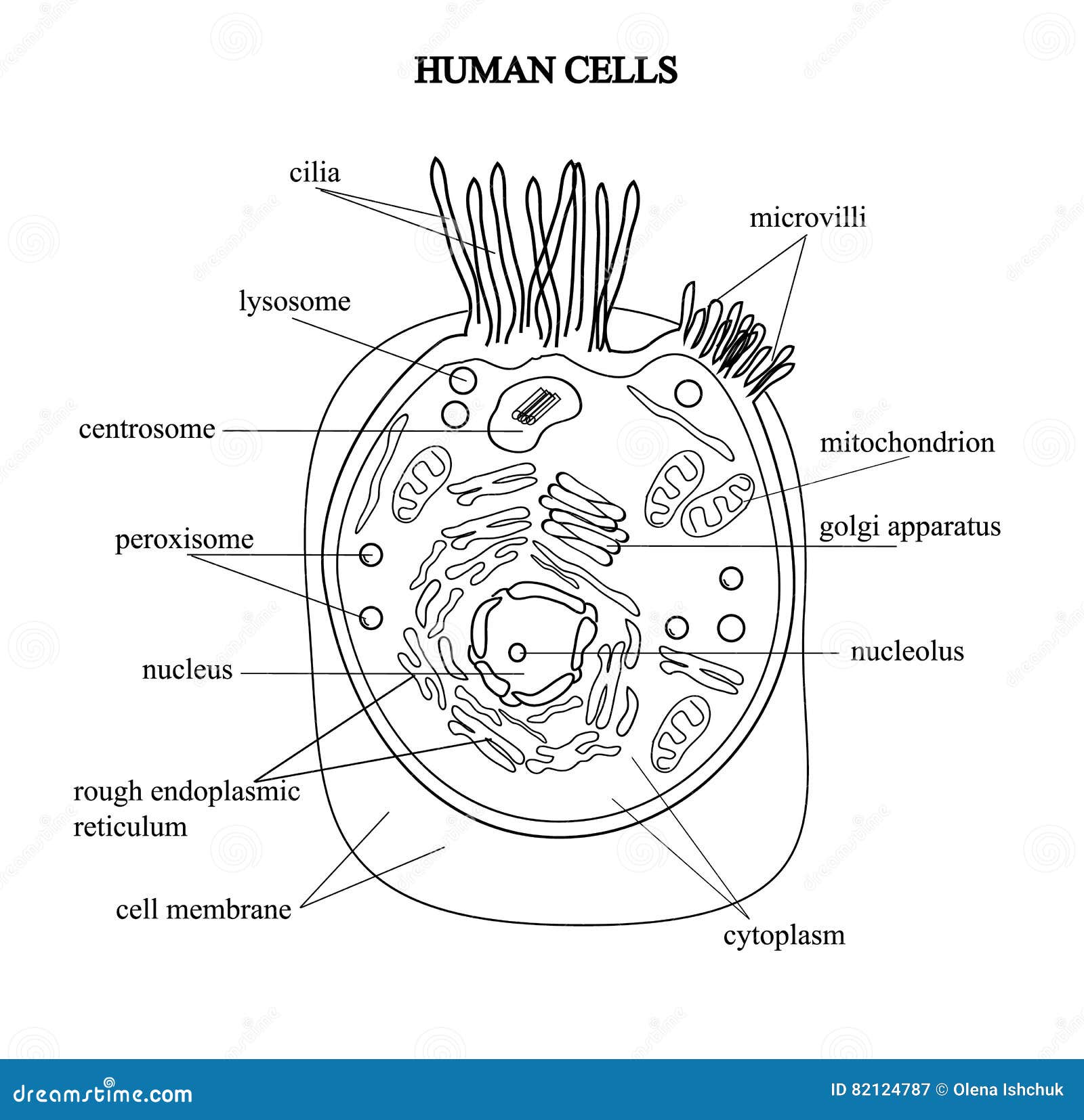
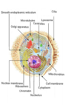
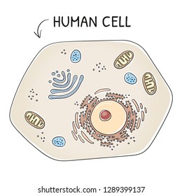
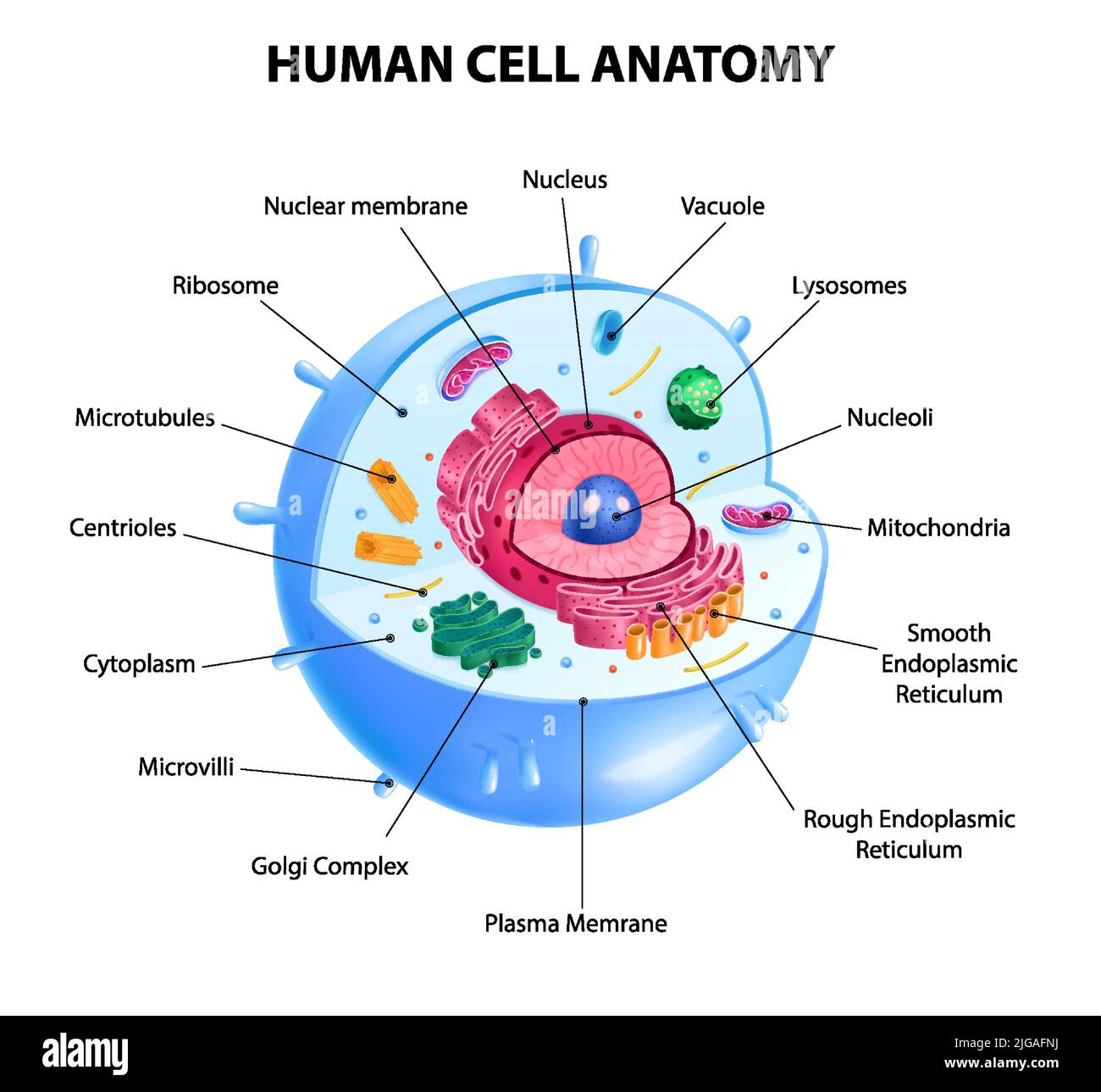
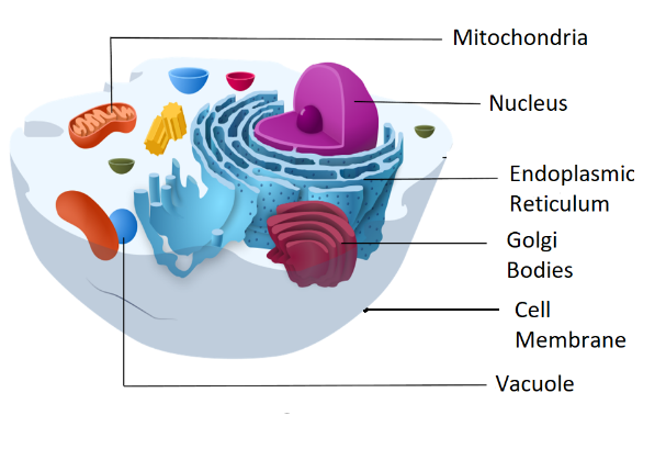
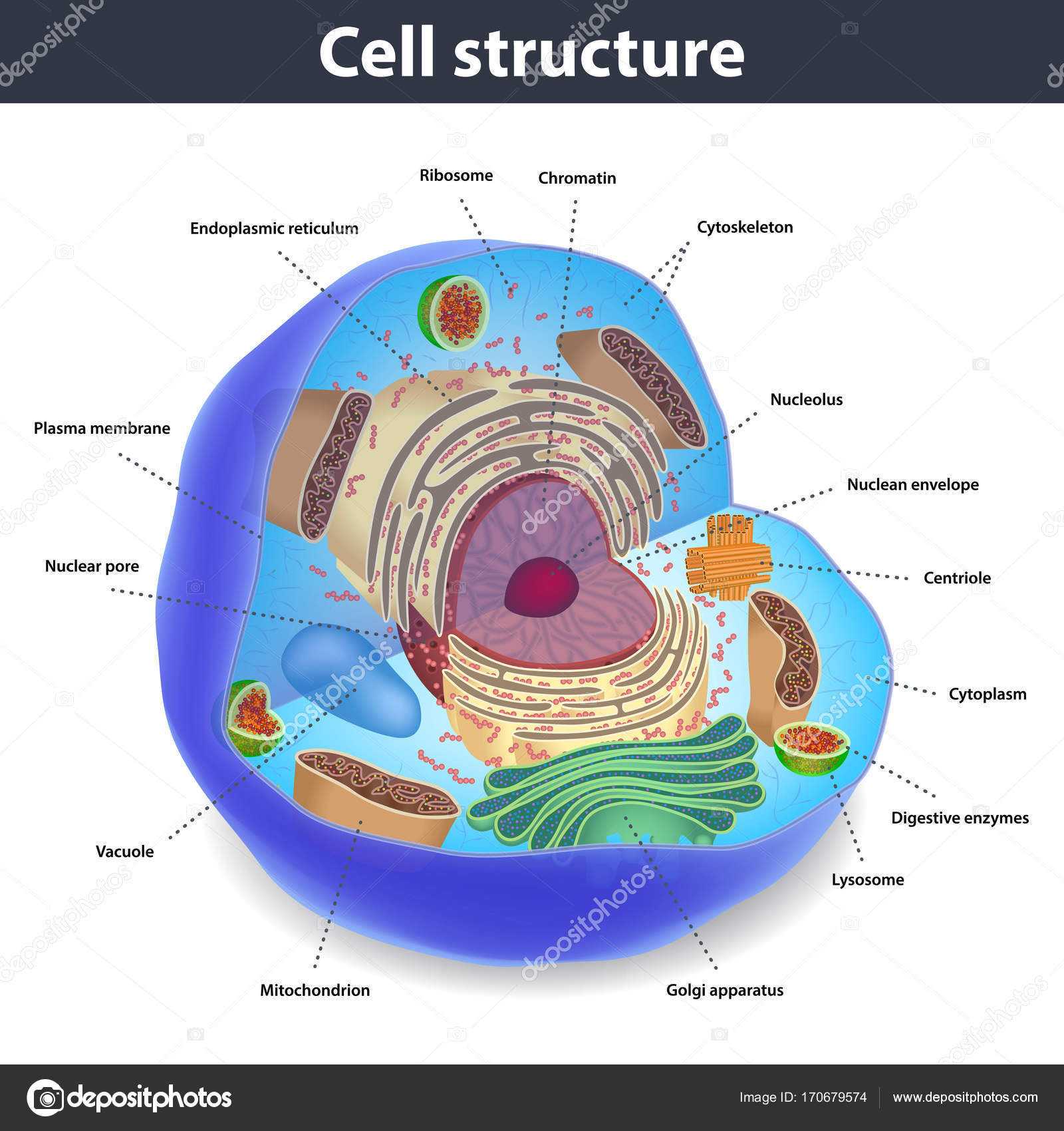
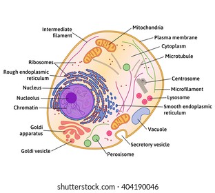
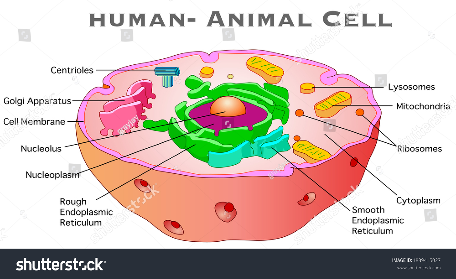
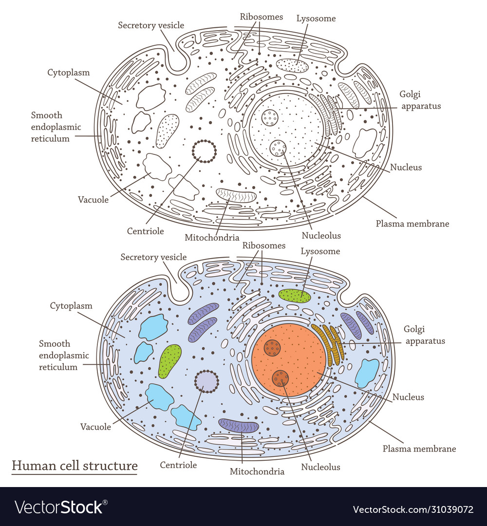
Human Cell Diagram, Parts, Pictures, Structure and Functions One of the few cells in the human body that lacks almost all organelles are the red blood cells. The main organelles are as follows : cell membrane; endoplasmic reticulum; Golgi apparatus; lysosomes; mitochondria; nucleus; perioxisomes; microfilaments and microtubules; Diagram of the human cell illustrating the different parts of the cell. Cell Membrane

Diagram of a human cell with labels
Draw a labelled diagram of human cells. - Careers360 Two solutions A and B each of 100L was made by dissolving 4g of NaOH and 9.8g of H2SO4 in water, respectively. The pH of the resultant solution obtained from mixing 40L of solution A and 10L of solution Q. The oxidation number of potassium in respectively is : Option: 1 Q. The relative strength of interionic/intermolecular forces in decreasing ... Labeled Diagrams of the Human Brain You'll Want to Copy Now All the functions are carried out without a single glitch and before you even bat an eyelid. The following are the different regions of the human brain and their functions. Labeled Diagrams of the Human Brain Central Core The central core consists of the thalamus, pons, cerebellum, reticular formation and medulla. A Well-labelled Diagram Of Animal Cell With Explanation - BYJUS Well-Labelled Diagram of Animal Cell The Cell Organelles are membrane-bound, present within the cells. There are various organelles present within the cell and are classified into three categories based on the presence or absence of membrane. Listed below are the Cell Organelles of an animal cell along with their functions.
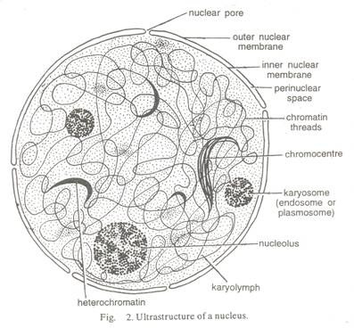
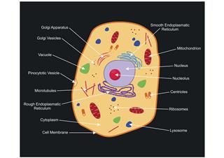
Diagram of a human cell with labels. File:Diagram human cell nucleus tr.svg - Wikimedia Commons Description: en: A diagram of a human cell nucleus, with Turkish labels. Translated version of File:Diagram human cell nucleus.svg, originally created and all rights released by Mariana Ruiz (User:LadyofHats).This image is also released to the public domain. az: İnsan hüceyrə nüvəsinin sxematik rəsmi, azərbaycanca yazılı. Source: File:Diagram human cell nucleus.svg PDF Human Cell Diagram, Parts, Pictures, Structure and Functions Diagram of the human cell illustrating the different parts of the cell. Cell Membrane The cell membraneis the outer coating of the cell and contains the cytoplasm, substances within it and the organelle. It is a double-layered membrane composed of proteins and lipids. Diagram of human skin structure — Science Learning Hub Diagram of human skin structure. Image. Add to collection. Tweet. Rights: University of Waikato Published 1 February 2011 Size: 100 KB Referencing Hub media. The epidermis is a tough coating formed from overlapping layers of dead skin cells. 03 Label the Cell Diagram | Quizlet Centrioles. Cell division. Cell Membrane. Outer boundary. Microtubules. Structure and support. Cytoplasm. Metabolic activities. Ribosome.
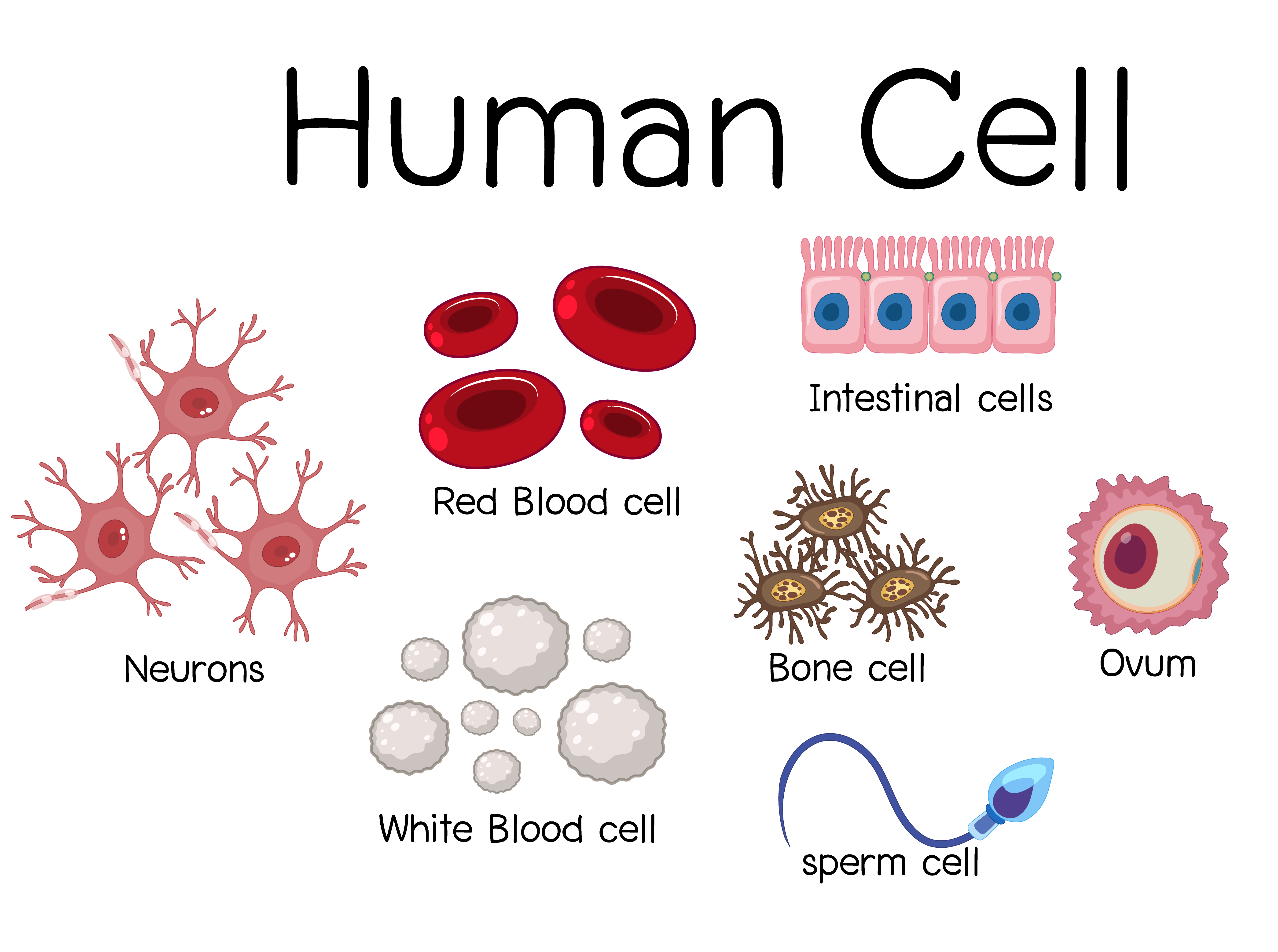
Human Cells for Kids - Worksheet, Cell Model Activity, Review Game Cells for Kids. As we began studying the human body we learned about the most basic unit of life - cells! This cells for kids lesson is filled with lots of engaging activities and cells printables for kids from kindergartners to elementary age students in grade 1, grade 2, grade 3, grade 4, grade 5, and grade 6. We used the following free printable All About Cells reader book to learn about ... A Labeled Diagram of the Plant Cell and Functions of its Organelles The cell membrane is a thin layer made up of proteins, lipids, and fats. It forms a protective wall around the organelles contained within the cell. It is selectively permeable and thus, regulates the transportation of materials needed for the survival of the organelles of the cell. Function: Protects the cell from its surroundings. Human Body Diagram - Bodytomy The human torso is also known as the 'trunk'. It is the central part of the body, and it is from here that the neck and the limbs extend. Some of the most critical human body organs are situated within the torso. The upper part consists of the heart and the lungs; these are protected by the rib cage. Anatomy and Physiology: Parts of a Human Cell - Visible Body Cells can be divided into four groups: somatic, gamete, germ, and stem. Somatic cells are all the cells in the body that aren't sex cells, like blood cells, neurons, and osteocytes. Gametes are sex cells that join together during sexual reproduction. Germ cells produce gametes.
A Labelled Diagram Of Neuron with Detailed Explanations - BYJUS A Labelled Diagram Of Neuron with Detailed Explanations Biology Biology Article Diagram Of Neuron Diagram Of Neuron A neuron is a specialized cell, primarily involved in transmitting information through electrical and chemical signals. They are found in the brain, spinal cord and the peripheral nerves. A neuron is also known as the nerve cell. Cells Diagram | Science Illustration Solutions - Edrawsoft Cells Diagram Symbols Edraw software offers you lots of symbols used in cells diagram like cell structure, paramecium, squamous cell, cell division, bacteria, cell membrane, eggs, sperm, zygote, an animal cell, SARS, tobacco mosaic, adenovirus, coliphage, herpesvirus, AIDS, pollen, plant cell model, onion tissue, etc. Cells Diagram Examples Human Cell Picture With Labels - Parts Of A Human Cell Diagram Of The ... Human Cell With Chinese Labels Full Color Panoramic Mug Spreadshirt from image.spreadshirtmedia.com This diagram depicts picture of the female body 744×992 with parts and labels. Rod and cone cells in the retina are photoreceptive cells which are able to detect visible light and convey this information to the brain.eyes signal information ... Interactive Cell Model - CELLS alive Cell Wall. Chloroplast. Smooth Endoplasmic Reticulum. Rough Endoplasmic Reticulum. Ribosomes. Cytoskeleton. RETURN to CELL DIAGRAM ...
Human Cell Structure Stock Illustration - Download Image Now ...
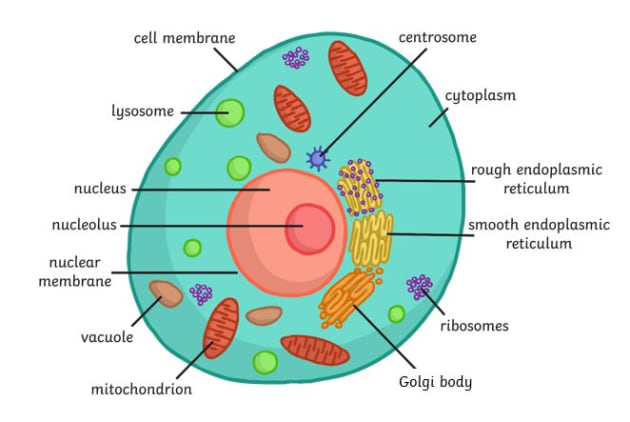
A Labeled Diagram of the Animal Cell and its Organelles A Labeled Diagram of the Animal Cell and its Organelles There are two types of cells - Prokaryotic and Eucaryotic. Eukaryotic cells are larger, more complex, and have evolved more recently than prokaryotes. Where, prokaryotes are just bacteria and archaea, eukaryotes are literally everything else.
Human Cell Structure Stock Illustrations – 29,816 Human Cell ...
Cell Organelles- Definition, Structure, Functions, Diagram Cell organelles are specialized entities present inside a particular type of cell that performs a specific function. There are various cell organelles, out of which, some are common in most types of cells like cell membranes, nucleus, and cytoplasm. However, some organelles are specific to one particular type of cell-like plastids and cell ...

4,442 Human Cell Diagram Stock Photos, Pictures & Royalty ...
Labeled diagram of the human kidney royalty-free images - Shutterstock Labeled diagram of the human kidney royalty-free images 189 labeled diagram of the human kidney stock photos, vectors, and illustrations are available royalty-free. See labeled diagram of the human kidney stock video clips Image type Orientation People Artists Sort by Popular Healthcare and Medical Anatomy Diseases, Viruses, and Disorders kidney
4,442 Human Cell Diagram Stock Photos, Pictures & Royalty ...
Labeled Plant Cell With Diagrams | Science Trends The parts of a plant cell include the cell wall, the cell membrane, the cytoskeleton or cytoplasm, the nucleus, the Golgi body, the mitochondria, the peroxisome's, the vacuoles, ribosomes, and the endoplasmic reticulum. Parts Of A Plant Cell The Cell Wall Let's start from the outside and work our way inwards.
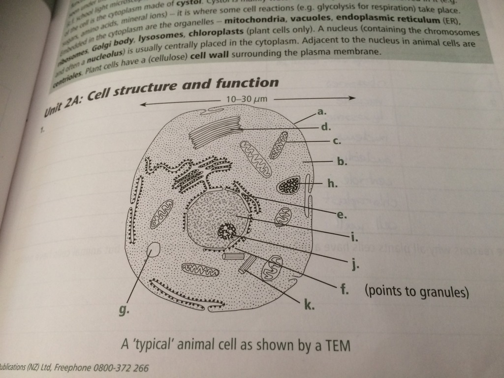
Solved] Make a model of a typical human cell with the major ...
Human Heart Diagram With Labels / Simple Diagram Of Human Heart With ... Diagram of the heart with labels. Diagram_of_the_human_heart_(no_labels).jpg (608 × 600 pixels, file size: ... Click on the diagram to show / hide labels. Cell structure and functions / animal cell vs plant cell / parts of cell / ch 8 science class 8 cbse . Diagram of the heart with labels.
What are cells in the human body? | Twinkl Teaching Wiki
Blank animal cell diagram to label human body anatomy. Blank animal cell diagram to label human body anatomy. 1100x1390 animal cell drawing labeled diagram of a typical animal cell, 1200x1200 animal cell drawing with labels animal cell diagrams to print. When drawing and labeling a diagram of the plasma membrane you should be sure to include. 0 ratings0% found this document useful (0 votes). 5th ...
Human cell diagram design 1338048 Vector Art at Vecteezy
Structure of Cell: Definition, Types, Diagram, Functions - Embibe What is a cell? Ans: The cell is the smallest, fundamental and functional unit that makes up all living beings including microorganisms, plants, animals and humans. Q.2. What are the five cell structures? Ans: A cell consists of many different structures that have definite shapes, structures, and functions of their own. Some of these structures are (1) Cell Wall (2) Mitochondria (3) Chloroplast (4) Cell Membrane and (5) Nucleus
Medibiz Tv | Articles
Cell: Structure and Functions (With Diagram) - Biology Discussion Eukaryotic Cells: 1. Eukaryotes are sophisticated cells with a well defined nucleus and cell organelles. 2. The cells are comparatively larger in size (10-100 μm). 3. Unicellular to multicellular in nature and evolved ~1 billion years ago. 4. The cell membrane is semipermeable and flexible. 5. These cells reproduce both asexually and sexually.
Human Cell Coloring And Labeling Page - Coloring Home
cell diagrams to label draw the diagrams of different types of cells and label them - Brainly.in. 18 Pictures about draw the diagrams of different types of cells and label them - Brainly.in : Image of an animal cell diagram with each organelle labeled | Célula animal, Proyecto célula, Blank Cell Diagrams | Biology | Pinterest | Plant cell, Plants and The o'jays and also Plant Cell Diagram And Label Simple - Cell ...

Pollock Human Cell Labels
Learn the parts of a cell with diagrams and cell quizzes For this exercise we'll start with an image of a cell diagram ready labeled. Study this and make sure that you're clear about which structure is found where. Cell diagram unlabeled It's time to label the cell yourself! As you fill in the cell structure worksheet, remember the functions of each part of the cell that you learned in the video.
Animal Cell Diagram by Science Source
Labelled Diagram Of A Human Cell Bone Cell Labeled Diagram Animal Cell ... Oct 7, 2018 - Labelled Diagram Of A Human Cell Bone Cell Labeled Diagram Animal Cell Free Printable To Label. ... Oct 7, 2018 - Labelled Diagram Of A Human Cell Bone Cell Labeled Diagram Animal Cell Free Printable To Label. Pinterest. Today. Explore. When autocomplete results are available use up and down arrows to review and enter to select ...
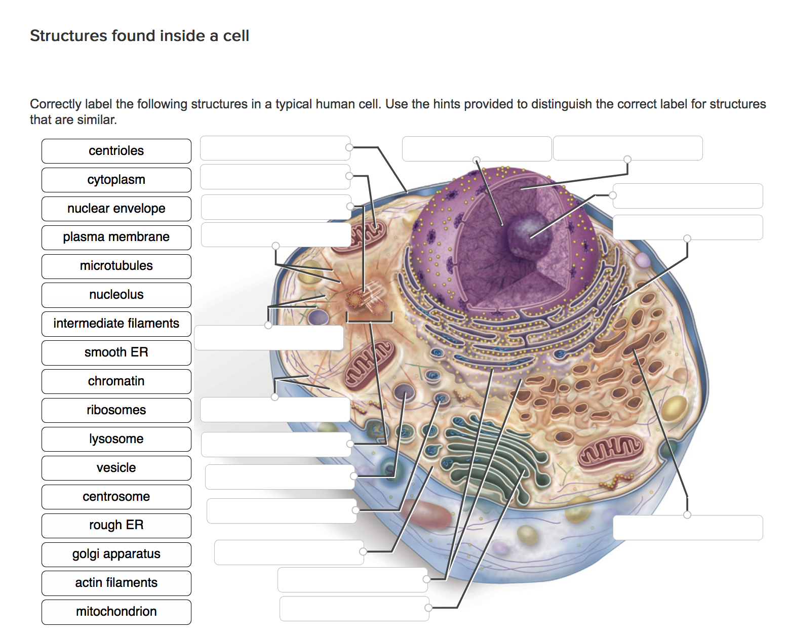
Solved Correctly label the following structures in a typical ...
A Well-labelled Diagram Of Animal Cell With Explanation - BYJUS Well-Labelled Diagram of Animal Cell The Cell Organelles are membrane-bound, present within the cells. There are various organelles present within the cell and are classified into three categories based on the presence or absence of membrane. Listed below are the Cell Organelles of an animal cell along with their functions.
Cell: Structure and Functions (With Diagram)
Labeled Diagrams of the Human Brain You'll Want to Copy Now All the functions are carried out without a single glitch and before you even bat an eyelid. The following are the different regions of the human brain and their functions. Labeled Diagrams of the Human Brain Central Core The central core consists of the thalamus, pons, cerebellum, reticular formation and medulla.

Education Chart Of Biology For Human Cell Diagram Royalty ...
Draw a labelled diagram of human cells. - Careers360 Two solutions A and B each of 100L was made by dissolving 4g of NaOH and 9.8g of H2SO4 in water, respectively. The pH of the resultant solution obtained from mixing 40L of solution A and 10L of solution Q. The oxidation number of potassium in respectively is : Option: 1 Q. The relative strength of interionic/intermolecular forces in decreasing ...
3,352 Cell organelles Images, Stock Photos & Vectors ...
Animal cell diagram hi-res stock photography and images - Alamy

What Is Going On Inside That Cell? | Human cell diagram, Cell ...

4,088 Eukaryotic Cell Stock Photos, Pictures & Royalty-Free ...
A Labeled Diagram of the Animal Cell and its Organelles ...

Animal Cell Structure Stock Illustration - Download Image Now ...

Human Cell Diagram, Parts, Pictures, Structure and Functions ...

Labelled Diagram Of A Human Cell Bone Cell Labeled Diagram ...
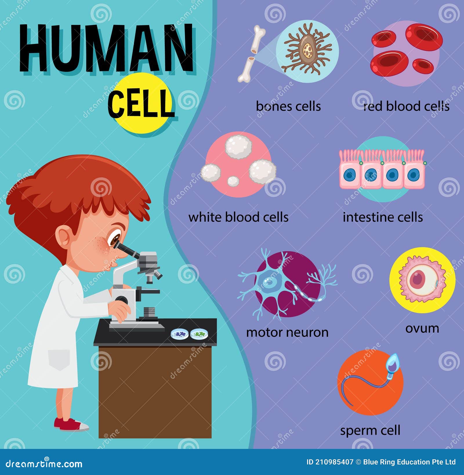
Diagram of Human Cell for Education Stock Vector ...
Realistic human cell anatomy diagram infographic poster ...
Draw a diagram of a typical cell and label the following ...

cell structure | Cell diagram, Animal cell drawing, Human ...
The structure of human cells, vector illustration Stock ...

Human Cell Diagram | Cell diagram, Human cell diagram, Plant ...
37,600 Cell diagram Images, Stock Photos & Vectors | Shutterstock
Human Cell Diagram (NCEA Level 2 Biology) Diagram | Quizlet
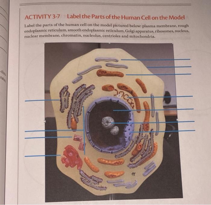
Solved ACTIVITY 3-7 Label the Parts of the Human Cell on the ...

Animal Cell Anatomy. Vector & Photo (Free Trial) | Bigstock
Animal Human Cell Diagram Structure Organelles Stock Vector ...
Human cell structure Royalty Free Vector Image

Human Physiology - Cell structure and function

Diagram of cell structure and function

Bio Geo Nerd: Cell Organelles | Cell diagram, Human cell ...
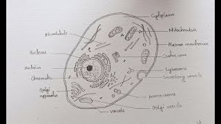
How to draw diagram of Animal Cell easily - step by step
Post a Comment for "38 diagram of a human cell with labels"