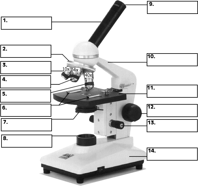
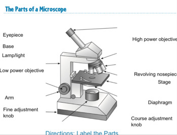
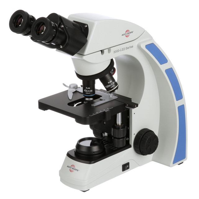
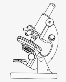
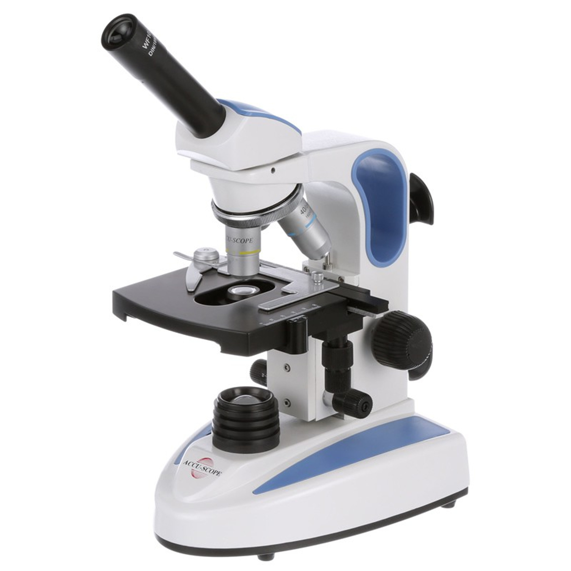
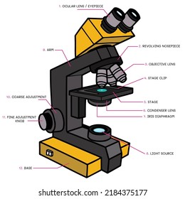
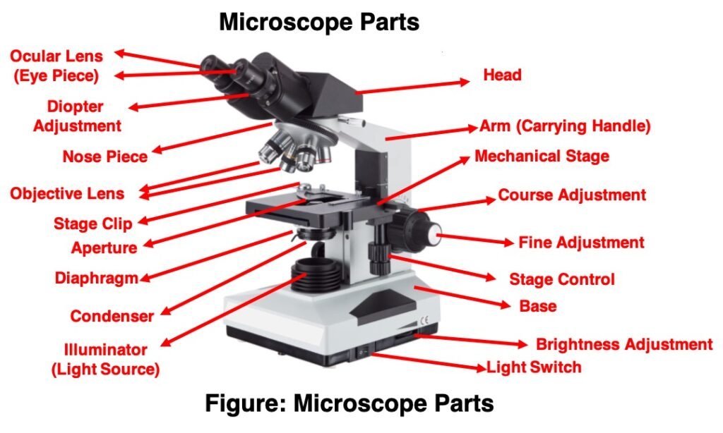
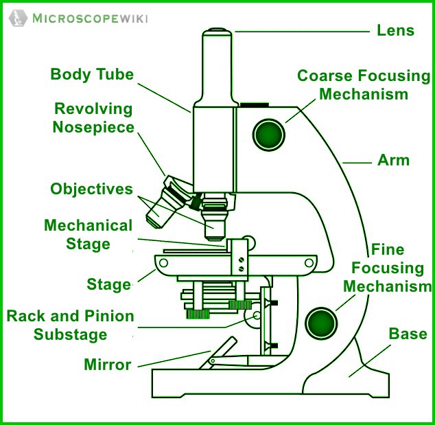
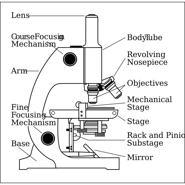
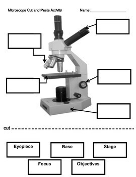
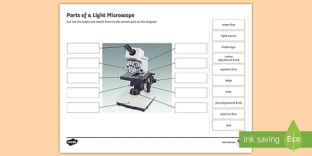
43 microscope diagram without labels
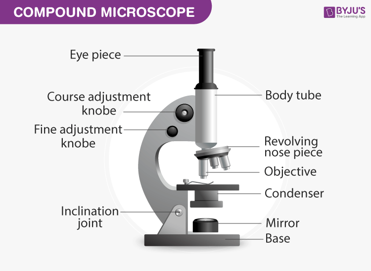
Parts of a microscope with functions and labeled diagram - Microbe Notes Figure: Diagram of parts of a microscope There are three structural parts of the microscope i.e. head, base, and arm. Head - This is also known as the body. It carries the optical parts in the upper part of the microscope. Base - It acts as microscopes support. It also carries microscopic illuminators. Looking at the Structure of Cells in the Microscope A typical animal cell is 10–20 μm in diameter, which is about one-fifth the size of the smallest particle visible to the naked eye. It was not until good light microscopes became available in the early part of the nineteenth century that all plant and animal tissues were discovered to be aggregates of individual cells. This discovery, proposed as the cell doctrine by Schleiden and …
Microscope Types (with labeled diagrams) and Functions Phase-contrast microscope labeled diagram Phase-contrast microscope functions: Its applications areas include In cases where the specimen is colorless and is very tiny In biology to conduct cellular level examination of microorganisms that can't be visualized using the bright field microscopy Interference Microscope

Microscope diagram without labels
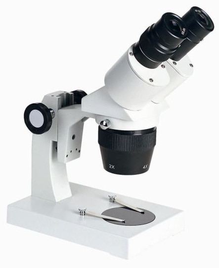
Simple Microscope - Parts, Functions, Diagram and Labelling Picture 1: The image above is a stereo microscope. Image source: made-in-china.com Picture 2: The image above is a confocal microscope. Image source:thorlabs.com Picture 3: The image above is parts of scanning electron microscope. Image source:britannica.com Picture 4: The picture is a transmission electron microscope. Image source: ysjournal.com en.wikipedia.org › wiki › Wikipedia:Citation_neededWikipedia:Citation needed - Wikipedia If someone tagged your contributions with a "Citation needed" tag or tags, and you disagree, discuss the matter on the article's talk page.The most constructive thing to do in most cases is probably to supply the reference(s) requested, even if you feel the tags are "overdone" or unnecessary. microscopeinternational.com › compound-microscopeCompound Microscope Parts, Functions, and Labeled Diagram Compound Microscope Definitions for Labels Eyepiece (ocular lens) with or without Pointer: The part that is looked through at the top of the compound microscope. Eyepieces typically have a magnification between 5x & 30x. Monocular or Binocular Head: Structural support that holds & connects the eyepieces to the objective lenses.
Microscope diagram without labels. PDF Parts of a Microscope Printables - Homeschool Creations Label the parts of the microscope. You can use the word bank below to fill in the blanks or cut and paste the words at the bottom. ... without needing to move the microscope ? the head •What is the magnification level on the eyepiece of a microscope?10x (see objective An Overview of Hyphae in Fungi, Their Function and Types. Jan 10, 2022 · In this alternative exercise, students will create a 3D annotated diagram of hyphae and its stages and types using food items or any other items easily accessible. Students must still use written ... Label the microscope — Science Learning Hub Label the microscope Interactive Add to collection Use this interactive to identify and label the main parts of a microscope. Drag and drop the text labels onto the microscope diagram. eye piece lens diaphragm or iris coarse focus adjustment stage base fine focus adjustment light source high-power objective Download Exercise Tweet Cambridge International AS amp A Level Biology Coursebook … 1.3 Plant and animal cells as seen with a light microscope 1.4 Measuring size and calculating magnification ... called ‘cells’. Each cell appeared to be an empty box surrounded by a wall. Hooke had discovered and described, without realising it, the fundamental unit of all living things. ev Cell structure ... Diagram of the electromagnetic ...
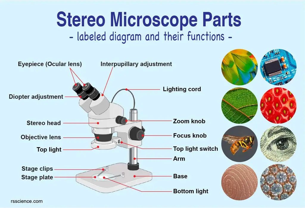
Microscope Diagram - Label Diagram | Quizlet The bottom of the microscope, used for support. ocular lens. Eyepiece of a microscope. Diaphragm. Regulates the amount of light on the specimen. nosepiece of microscope. holds the objective lenses. objective lens. The lens on a light microscope that is closest to the stage. Free Microscope Worksheets for Simple Science Fun for Your Students 1. Parts of a Microscope . The first worksheet labels the different parts of a microscope, including the base, slide holder, and condenser. If you have a microscope, compare and contrast this worksheet to it.Also, your kids can color this microscope diagram in and read the words to each part of the microscope. Microscope, Microscope Parts, Labeled Diagram, and Functions The Microscopes parts divided into three different structural parts Head, Base, and Arms. Head/Body: It contain the optical parts in the upper part of the microscope. Arm: It supports the tube and connects it to the base. Base: The bottom of the microscope, used for support. Optical Components of Microscope rsscience.com › stereo-microscopeParts of Stereo Microscope (Dissecting microscope) – labeled ... Labeled part diagram of a stereo microscope Major structural parts of a stereo microscope. There are three major structural parts of a stereo microscope. The viewing Head includes the upper part of the microscope, which houses the most critical optical components, including the eyepiece, objective lens, and light source of the microscope.
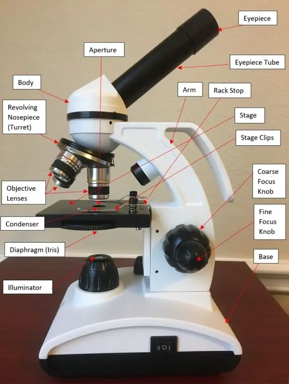
Download Label The Microscope Diagram - Robot | Transparent PNG ... Label The Microscope Diagram - Robot. You may also like PNG. Label The Microscope Diagram - Robot. 850*896. 0. 0. PNG. Nervous System Diagram Arrows - Nervous System Diagram Without Labels. 541*1023. 0. 0. PNG. Svg Diagram At Getdrawings Com Free For Personal - Diagram Of The Heart No Labels. 546*678. 0. 0. PNG. Human Figure Png - Human Body ... A Study of the Microscope and its Functions With a Labeled Diagram ... Here, unlabeled microscope diagrams have been provided for your perusal, which will help you practice and test your understanding of the instrument. Types of Microscopes Depending on the source of illumination, microscopes can be divided into two categories. They are: autospurgo.roma.itRefrigerator Flashing Whirlpool Lights Led Search: Whirlpool Refrigerator Led Lights Flashing. After switching to LEDs or when replacing a faulty LED lamp in some cases the LED light will start flickering We will explain temperature settings, alarm sounds, door not closing, water filter changes, not cooling issues, not making ice, no power, strange sounds, leveling, ice makers, water dispensers This refrigerator has the icemaker & bin ... 16 Parts of a Compound Microscope: Diagrams and Video The 16 core parts of a compound microscope are: Head (Body) Arm Base Eyepiece Eyepiece tube Objective lenses Revolving Nosepiece (Turret) Rack stop Coarse adjustment knobs Fine adjustment knobs Stage Stage clips Aperture Illuminator Condenser Diaphragm Video: Parts of a compound Microscope with Diagram Explained
› articles › 10Frontiers | A Comprehensive Review of Fluorescence ... Apr 12, 2021 · Fluorescence correlation spectroscopy (FCS) is a powerful technique for quantification of molecular dynamics, and it has been widely applied in diverse fields, e.g., biomedicine, biophysics, and chemistry. By time-correlation of the fluorescence fluctuations induced by molecules diffusing through a focused light, FCS can quantitatively evaluate the concentration, diffusion coefficient, and ...
Microscope Parts, Function, & Labeled Diagram - slidingmotion Microscope Parts Labeled Diagram The principle of the Microscope gives you an exact reason to use it. It works on the 3 principles. Magnification Resolving Power Numerical Aperture. Parts of Microscope Head Base Arm Eyepiece Lens Eyepiece Tube Objective Lenses Nose Piece Adjustment Knobs Stage Aperture Microscopic Illuminator Condenser Lens
Microscope Objective Lens | Products | Leica Microsystems The objective lens is a critical part of the microscope optics. The microscope objective is positioned near the sample, specimen, or object being observed. It has a very important role in imaging, as it forms the first magnified image of the sample. The numerical aperture (NA) of the objective indicates its ability to gather light and largely determines the microscope’s …
Fluorescence Resonance Energy Transfer (FRET) Microscopy Presented in Figure 3 is a Jablonski diagram illustrating the coupled transitions involved between the donor emission and acceptor absorbance in fluorescence resonance energy transfer. Absorption and emission transitions are represented by straight vertical arrows (green and red, respectively), while vibrational relaxation is indicated by wavy ...
Compound Microscope Parts - Labeled Diagram and their Functions The eyepiece (or ocular lens) is the lens part at the top of a microscope that the viewer looks through. The standard eyepiece has a magnification of 10x. You may exchange with an optional eyepiece ranging from 5x - 30x. [In this figure] The structure inside an eyepiece. The current design of the eyepiece is no longer a single convex lens.
Label The Microscope Diagram - Robot PNG Image | Transparent PNG Free ... Label The Microscope Diagram - Robot is a high-resolution transparent PNG image. It is a very clean transparent background image and its resolution is 850x896 , please mark the image source when quoting it.
Parts of Stereo Microscope (Dissecting microscope) – labeled diagram ... Labeled part diagram of a stereo microscope ... (based on color bands and their respective labels), the objectives of a dissecting microscope are located in a cylindrical cone and, therefore, are not directly seen. ... but large enough to be seen or handled without the aid of a high power compound microscope. Thus, stereo microscopes have a ...
en.wikipedia.org › wiki › Electron_microscopeElectron microscope - Wikipedia An electron microscope is a microscope that uses a beam of accelerated electrons as a source of illumination. As the wavelength of an electron can be up to 100,000 times shorter than that of visible light photons , electron microscopes have a higher resolving power than light microscopes and can reveal the structure of smaller objects.
Fluorescence In Situ Hybridization (FISH) | Learn Science at Scitable Cytogenetics entered the molecular era with the introduction of in situ hybridization, a procedure that allows researchers to locate the positions of specific DNA sequences on chromosomes. Since ...
Electron microscope - Wikipedia An electron microscope is a microscope that uses a beam of accelerated electrons as a source of illumination. As the wavelength of an electron can be up to 100,000 times shorter than that of visible light photons, electron microscopes have a higher resolving power than light microscopes and can reveal the structure of smaller objects.. Electron microscopes use shaped magnetic …
Label the microscope Diagram | Quizlet Diaphragm. Regulates the amount of light on the specimen. Light Source. Projects light upwards through the diaphragm, the specimen, and the lenses. Arm. supports the body tube. Stage. Supports the slide being viewed. Coarse Adjustment.
List of Top 7 Types of Microscopes (With Diagram) - Biology Discussion ADVERTISEMENTS: List of top seven types of microscopes:- 1. Phase Contrast Microscope 2. Interference Contrast Microscope 3. Ultraviolet Microscope 4. Fluorescence Microscope 5. Immunofluorescence 6. Dark-Field Microscope 7. Electron Microscope. Type # 1. Phase Contrast Microscope: This microscope was developed by Fritz Zernikes (1935), a Dutch physicist who was awarded Nobel Prize in 1953 ...
Microorganisms: Friend and Foe Class 8 Extra Questions Oct 11, 2019 · Pull out a gram or bean plant from the field. Observe its roots. You will find round struc¬tures called root nodules on the roots. Draw a diagram of the root and show the root nod¬ules. Answer: Question 2. Collect the labels from the bottles of jams and jellie on the labels. Answer: Do it yourself. Question 3. Visit a dcotor.
Labeling the Parts of the Microscope | Microscope World Resources Labeling the Parts of the Microscope This activity has been designed for use in homes and schools. Each microscope layout (both blank and the version with answers) are available as PDF downloads. You can view a more in-depth review of each part of the microscope here. Download the Label the Parts of the Microscope PDF printable version here.
› products › microscopeMicroscope Objective Lens | Products | Leica Microsystems The objective lens is a critical part of the microscope optics. The microscope objective is positioned near the sample, specimen, or object being observed. It has a very important role in imaging, as it forms the first magnified image of the sample. The numerical aperture (NA) of the objective indicates its ability to gather light and largely determines the microscope’s resolution, the ...
Free Microscope Worksheets for Simple Science Fun for Your Students The Microscope includes a colorful drawing of a microscope and lines to add observations and such. Microscope Lab Report includes colorful drawings of a microscope, test tubes, and slides. It also has 2 circles (to draw observations) and lines for writing. Get Your Free Set of Microscope Worksheets! YES!
Simple Microscope - Diagram (Parts labelled), Principle, Formula and Uses The working principle of a simple microscope is that when a lens is held close to the eye, a virtual, magnified and erect image of a specimen is formed at the least possible distance from which a human eye can discern objects clearly. Magnification formula The magnification power of a simple microscope is expressed as: M = 1 + D/F Where
microscopeinternational.com › compound-microscopeCompound Microscope Parts, Functions, and Labeled Diagram Compound Microscope Definitions for Labels Eyepiece (ocular lens) with or without Pointer: The part that is looked through at the top of the compound microscope. Eyepieces typically have a magnification between 5x & 30x. Monocular or Binocular Head: Structural support that holds & connects the eyepieces to the objective lenses.
en.wikipedia.org › wiki › Wikipedia:Citation_neededWikipedia:Citation needed - Wikipedia If someone tagged your contributions with a "Citation needed" tag or tags, and you disagree, discuss the matter on the article's talk page.The most constructive thing to do in most cases is probably to supply the reference(s) requested, even if you feel the tags are "overdone" or unnecessary.
Simple Microscope - Parts, Functions, Diagram and Labelling Picture 1: The image above is a stereo microscope. Image source: made-in-china.com Picture 2: The image above is a confocal microscope. Image source:thorlabs.com Picture 3: The image above is parts of scanning electron microscope. Image source:britannica.com Picture 4: The picture is a transmission electron microscope. Image source: ysjournal.com










Post a Comment for "43 microscope diagram without labels"